Microscopy and Image Analysis Core Facility
Weill Cornell Medicine
Overview of services offered at our core facility
Please feel free to contact us using this form
Some images collected at our core facility
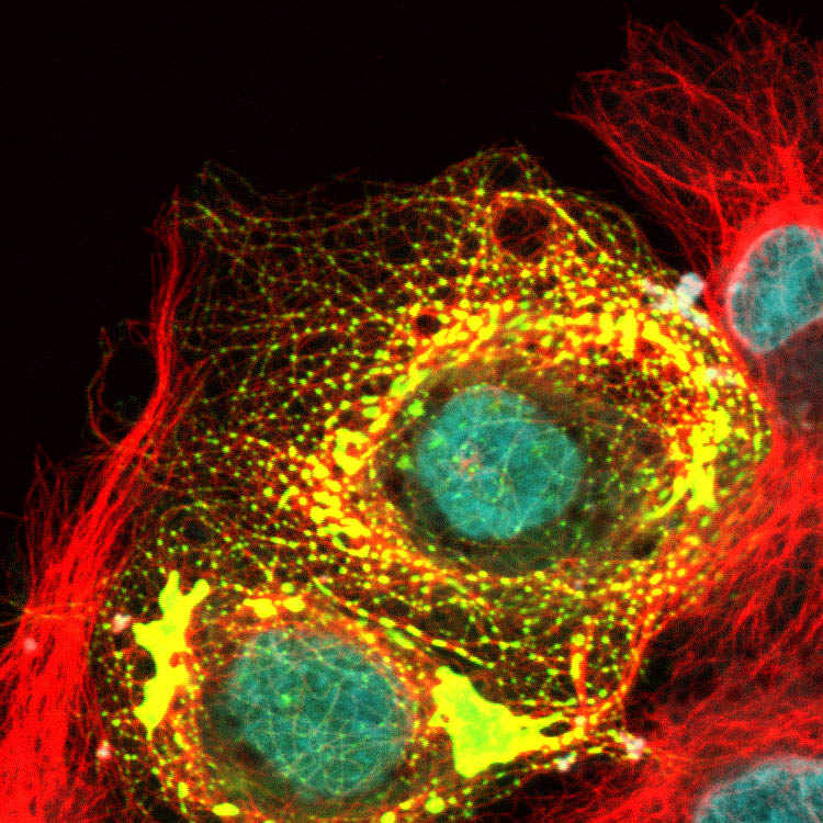


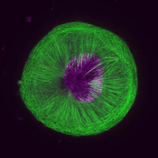


Credit: Katsuhiro Kita, Paraskevi Giannakakou (WCM)
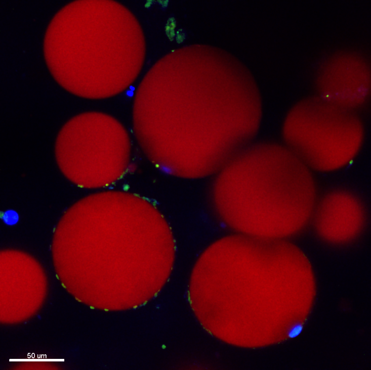
Credit: Manu Jain, Sushmita Mukherjee (WCM)
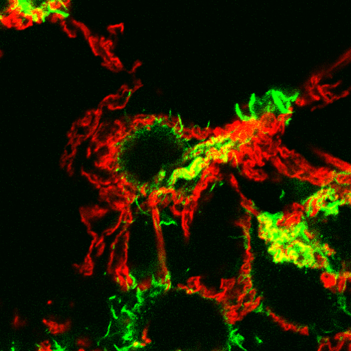
Credit: Priya Bharadwaj, Kristy Brown (WCM)
Credit: Katsuhiro Kita, Paraskevi Giannakakou (WCM)
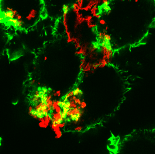
Credit: Inna Grosheva, Frederick Maxfield (WCM)
The yellow color indicates places where cholesterol has entered an organelle into which the transferrin has been delivered.
Credit: Mingming Hao, Frederick Maxfield (WCM)
Credit: Inna Grosheva, Frederick Maxfield
Credit: Amitabha Majumdar, Frederick Maxfield (WCM)
Credit: Katsuhiro Kita, Paraskevi Giannakakou (WCM)
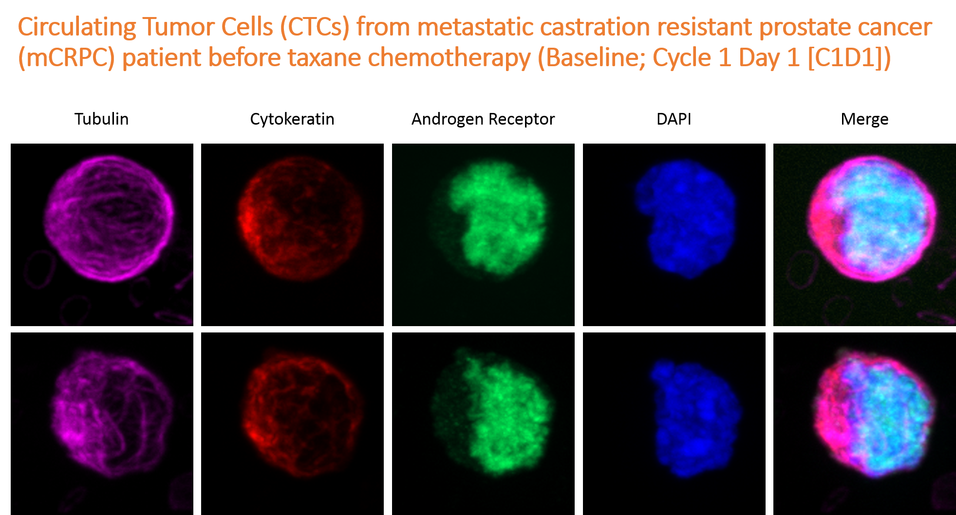
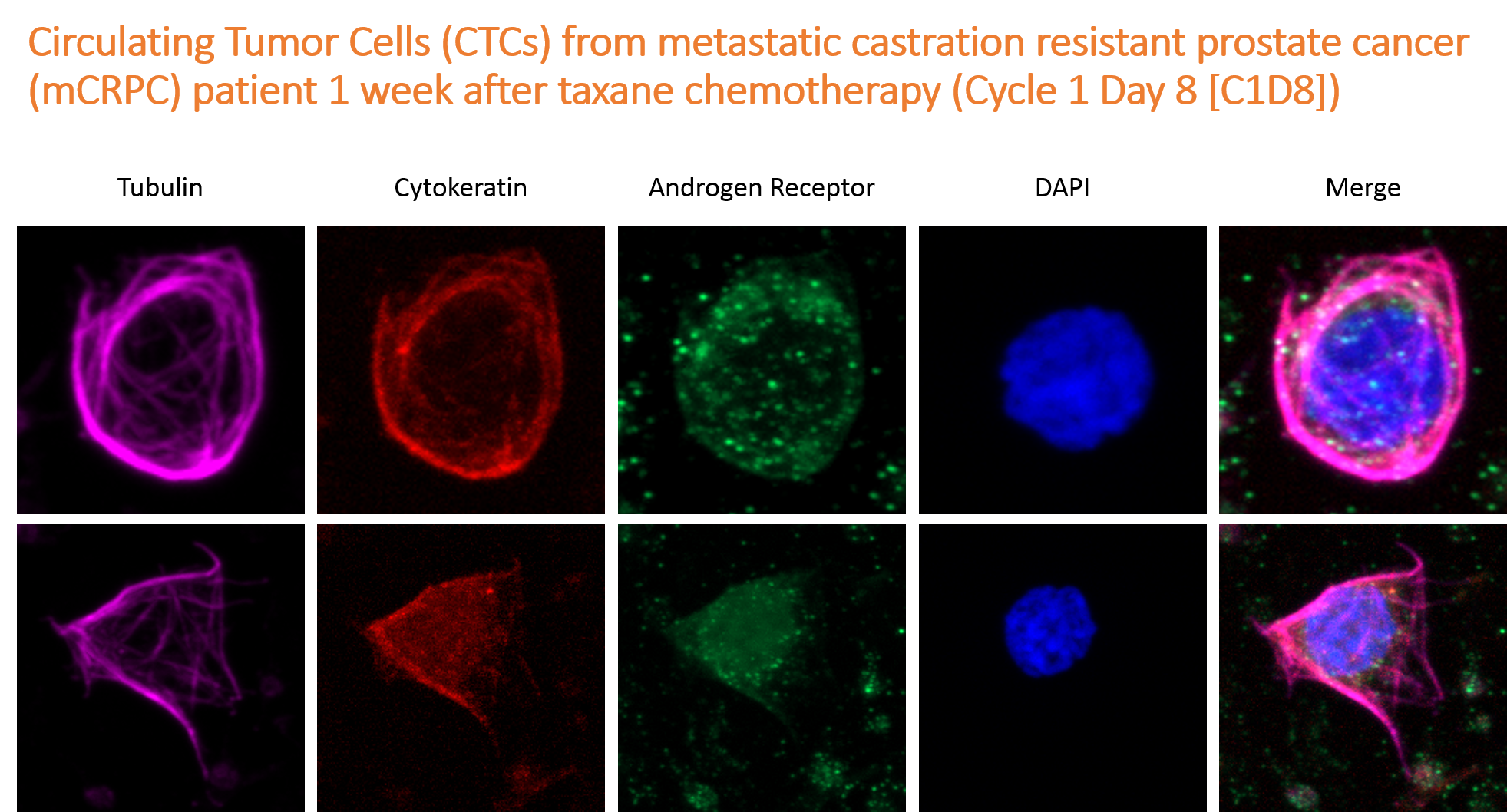
Credit: David Worroll, Paraskevi Giannakakou (WCM)
Credit: David Worroll, Paraskevi Giannakakou (WCM)