Confocal microscopy
Zeiss LSM 880
(inverted configuration)
Features include:
Zeiss LSM880 confocal microscope
Fluorescence confocal, Brightfield imaging and data capture
Inverted AxioObserver microscope stand
Computer controlled by ZEN (Zeiss Enhanced Navigation) software
Laser excitations at 405, 458, 488, 514, 561, 594 and 633 nm
Available lenses: 10x, 20x, 25x, 40x and 63x (All DIC)
X-Y Motorized stage for tiling and multiple region imaging
High sensitivity GAsP detector
Dedicated “blue” and “red” PMTs for the low and high end of the spectrum
Highly efficient light path
Simultaneous Fluorescence and DIC imaging
Experimental Designer for multi-dimensional acquisition
Tiling Macro for large image regions
Zeiss LSM 880 with AiryScan, FAST Airyscan and 32-channel GaAsP detector array for spectral imaging
(inverted configuration)
Zeiss LSM880 with Airyscan and environmental chamber for live cell imaging
Features include:
Fluorescence confocal, Brightfield imaging and data capture
Inverted AxioObserver microscope stand
Computer controlled by ZEN (Zeiss Enhanced Navigation) software
Laser excitations at 405, 458, 488, 514, 561, 594 and 633 nm
Available lenses: 10x, 20x, 25x, 40x, 63x (All DIC)
X-Y Motorized stage for tiling and multiple region imaging
Incubation chamber and heating inserts with CO2 for live cell imaging
High sensitivity 32 channel GAsP detector for visible spectrum detection and linear unmixing
Dedicated “blue” and cooled “red” PMTs for the low and high end of the spectrum
Highly efficient light path
Simultaneous Fluorescence and DIC imaging in confocal mode
Experimental Designer for multi-dimensional acquisition
Tiling Macro for large image regions
AIRYSCAN and FAST AIRYSCAN high resolution GaAsP detectors
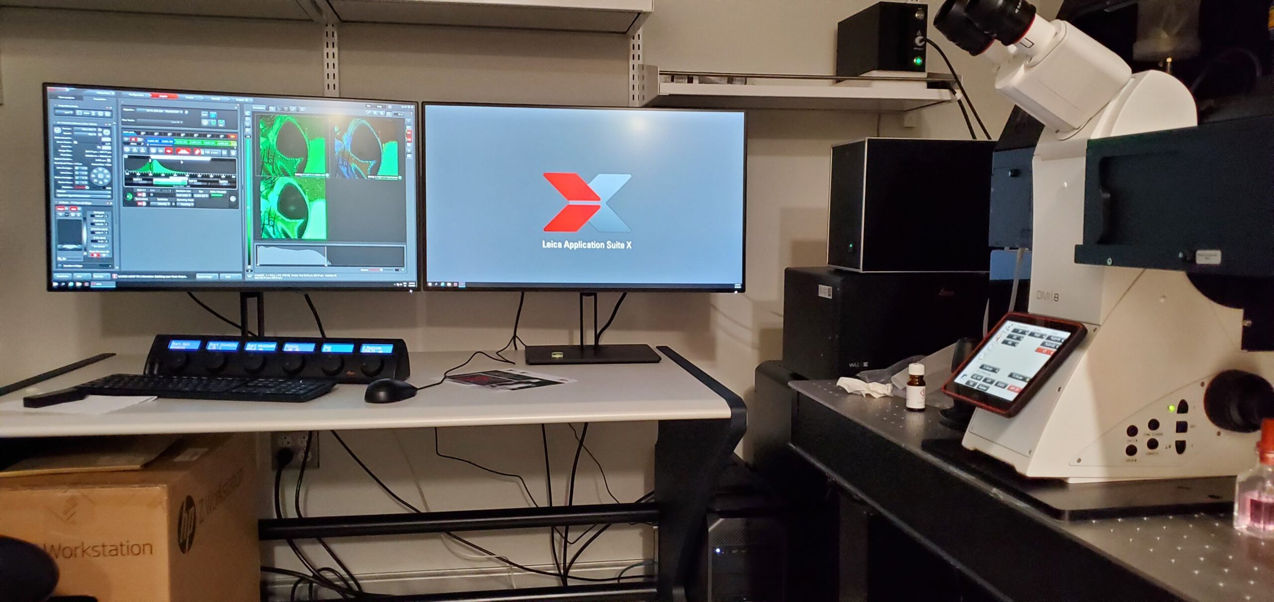
Leica Stellaris 8 with FALCON (FAst Lifetime CONtrast) and tunable White Light Laser illumination
(inverted configuration)
Features include:
White Light Laser (440nm-795nm emissions)
FALCON TauSense technology for Fast Lifetime Imaging
5 High sensitivity HyD confocal fluorescence detectors
FRET, FRAP: quantify fast cellular processes, like molecular interaction or molecular environment changes
Solid state 405 nm laser
Transmitted light detector
Resonant and non-resonant scanning modes for optimal scanning speeds
Motorized stage for X-Y tiling and Z-stack collection
Automatic Focus Control (AFC)
Fluorescence confocal, Brighfield imaging and data collection
Inverted DMi 8 Premium stand
LASX Software
Laser excitations: White Light laser (tunable from 440 - 790 nm); solid state 405 nm laser
Objectives: CS2 /low background lenses: 5x, 10x, 20x,40x, 63x-Oil-DIC, 63x-Glyc-DIC (four dry lenses 5 - 40x allow multi-position time-lapse imaging with more ease)
Leica SP8 with DIVE optics (combined confocal and multiphoton)
Leica SP8 with DIVE optics, combined confocal and multiphoton microscope
(upright configuration)
Features include:
3 confocal (descanned) detectors - two HyD and one PMT
"Filterless" acquisition (custom emission bandwidths)
“Navigator” for mark and find, and tile scanning
Switchable between resonant and non-resonant mode for optimal scanning speeds and resolution
Adjustable height fixed stage, along with a smaller motorized XY stage
Laser excitations: 405, 458, 488, 522, 638 nm.
Objectives: 5x Air, 10x Air, 20x Air, 40x Oil, 63x Oil Immersion. All DIC for simultaneous transmitted light imaging.
For the multiphoton options on this instrument, please click the button below.
ImageXpress Micro Confocal Imager (automated spinning disk confocal)
(inverted configuration)
Features include:
ImageXpress Micro Confocal (spinning disk) automated imager
Automated imaging of fixed or live cells in multi-well plates or glass slides
Widefield imaging or spinning disk confocal imaging with 42µm and 60µm pinholes
Solid-state light engine and filter sets for DAPI, FITC, TRITC, TxRed and Cy5 fluorescence
High quantum efficiency 16-bit sCMOS camera enabling >3 log dynamic range and a large field of view (1.96 mm2 at 10X)
Long working-distance objectives ranging from 2X to 40X
Transmitted light and Phase Contrast imaging for non-fluorescent samples
Multi-day, time-lapse imaging with temperature, humidity and CO2 control for live cells
Unattended screening of up to 45 fixed plates per run
Automated, fully customizable 2D and 3D image analysis on our MetaXpress workstations. Images can be exported for analysis in other packages
Sample prep facilitated by two MultiDrop384 dispensers and a BioTek plate washer


