Credit: Katsuhiro Kita, Paraskevi Giannakakou (WCM)

Credit: Manu Jain, Sushmita Mukherjee (WCM)

Credit: Priya Bharadwaj, Kristy Brown (WCM)

Credit: Katsuhiro Kita, Paraskevi Giannakakou (WCM)

Credit: Inna Grosheva, Frederick Maxfield (WCM)

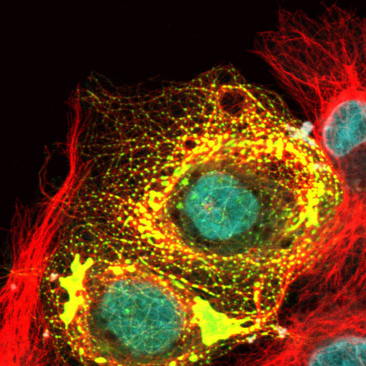
The yellow color indicates places where cholesterol has entered an organelle into which the transferrin has been delivered.
Credit: Mingming Hao, Frederick Maxfield (WCM)

Credit: Inna Grosheva, Frederick Maxfield

Credit: Amitabha Majumdar, Frederick Maxfield (WCM)

Credit: Katsuhiro Kita, Paraskevi Giannakakou (WCM)

Credit: David Worroll, Paraskevi Giannakakou (WCM)

Credit: David Worroll, Paraskevi Giannakakou (WCM)


Credit: Katsuhiro Kita, Paraskevi Giannakakou (WCM)
Credit: Manu Jain, Sushmita Mukherjee (WCM)
Credit: Priya Bharadwaj, Kristy Brown (WCM)
Credit: Katsuhiro Kita, Paraskevi Giannakakou (WCM)
Credit: Inna Grosheva, Frederick Maxfield (WCM)
The yellow color indicates places where cholesterol has entered an organelle into which the transferrin has been delivered.
Credit: Mingming Hao, Frederick Maxfield (WCM)
Credit: Inna Grosheva, Frederick Maxfield
Credit: Amitabha Majumdar, Frederick Maxfield (WCM)
Credit: Katsuhiro Kita, Paraskevi Giannakakou (WCM)
Credit: David Worroll, Paraskevi Giannakakou (WCM)
Credit: David Worroll, Paraskevi Giannakakou (WCM)
