
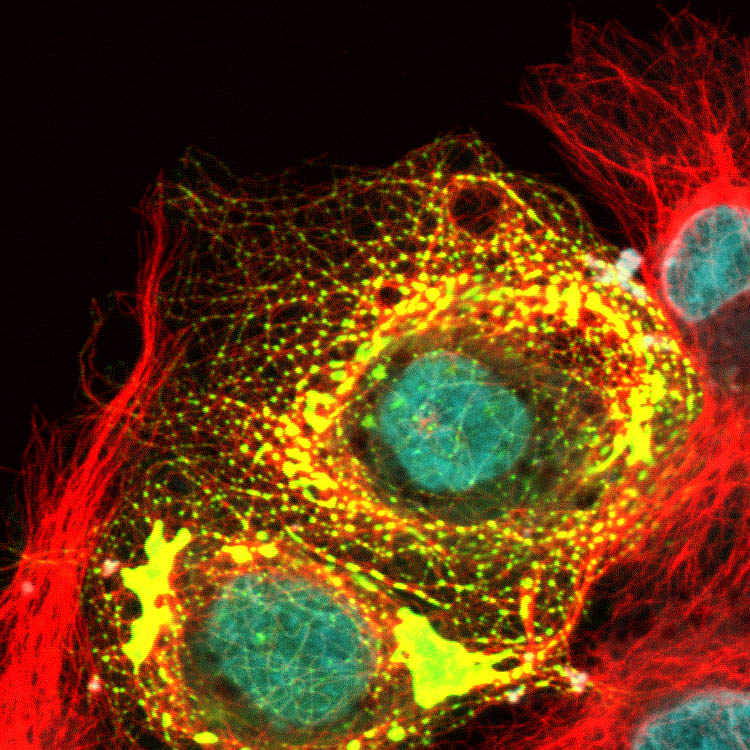
COS-7 cells expressing abnormal form of microtubule plus-end binding protein. MTs = red, yellow = plus-end binding protein
Credit: Katsuhiro Kita, Paraskevi Giannakakou (WCM)

Fresh mouse colon (unfixed, unstained) imaged using multiphoton microscopy. Green and Blue: autofluorescence at short and long wavelengths; red: Second Harmonic Generation (SHG)
Credit: Manu Jain, Sushmita Mukherjee (WCM)
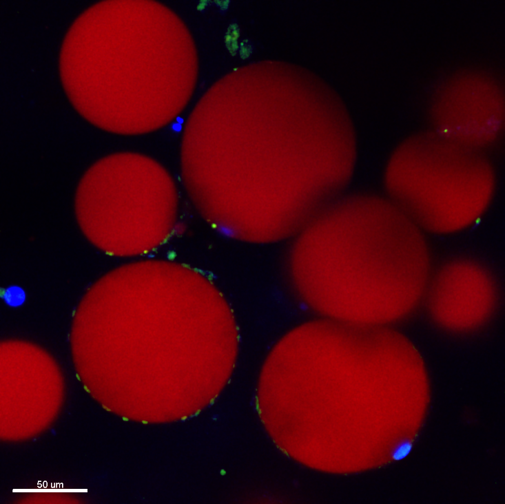
Primary human adipocytes isolated from breast tissue were stained with lipidTox (red)
Credit: Priya Bharadwaj, Kristy Brown (WCM)

Gastric cancer cell stained with endogenous MT-end binding protein and MTs. MTs = green, red = end-binding protein
Credit: Katsuhiro Kita, Paraskevi Giannakakou (WCM)
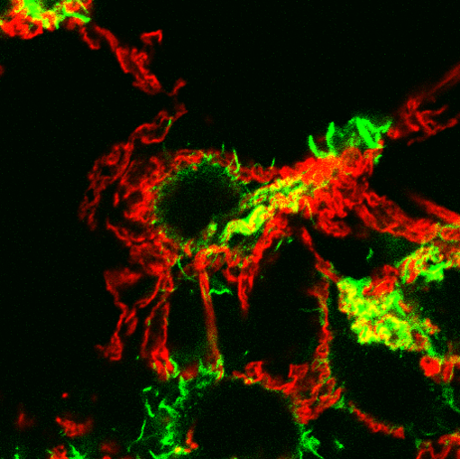
Lipoproteins (red) being digested by macrophages (green)
Credit: Inna Grosheva, Frederick Maxfield (WCM)

Two living cells showing fluorescent sterol (green), a fluorescent lipid (blue) and an iron-carrying protein (transferrin, red)
The yellow color indicates places where cholesterol has entered an organelle into which the transferrin has been delivered.
Credit: Mingming Hao, Frederick Maxfield (WCM)
Lipoproteins (red) being digested by macrophages (green)
Credit: Inna Grosheva, Frederick Maxfield

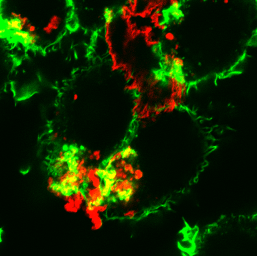
A microglial cell (green) has internalized Alzheimer’s amyloid protein (red)
Credit: Amitabha Majumdar, Frederick Maxfield (WCM)
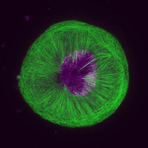
Gastric cancer cell treated with a tool compound showed a round and flat phenotype with strange MT bundles. MTs = green, purple = DAPI
Credit: Katsuhiro Kita, Paraskevi Giannakakou (WCM)
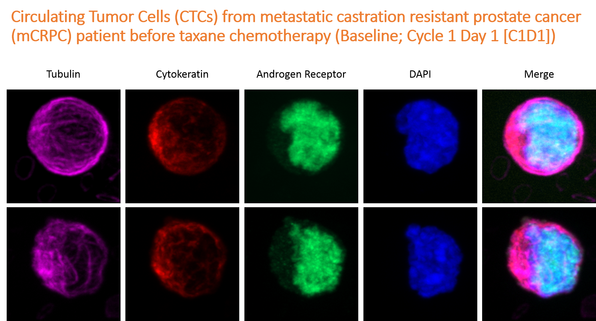
Circulating Tumor Cells (CTCs) pre-treatment with taxane
Credit: David Worroll, Paraskevi Giannakakou (WCM)
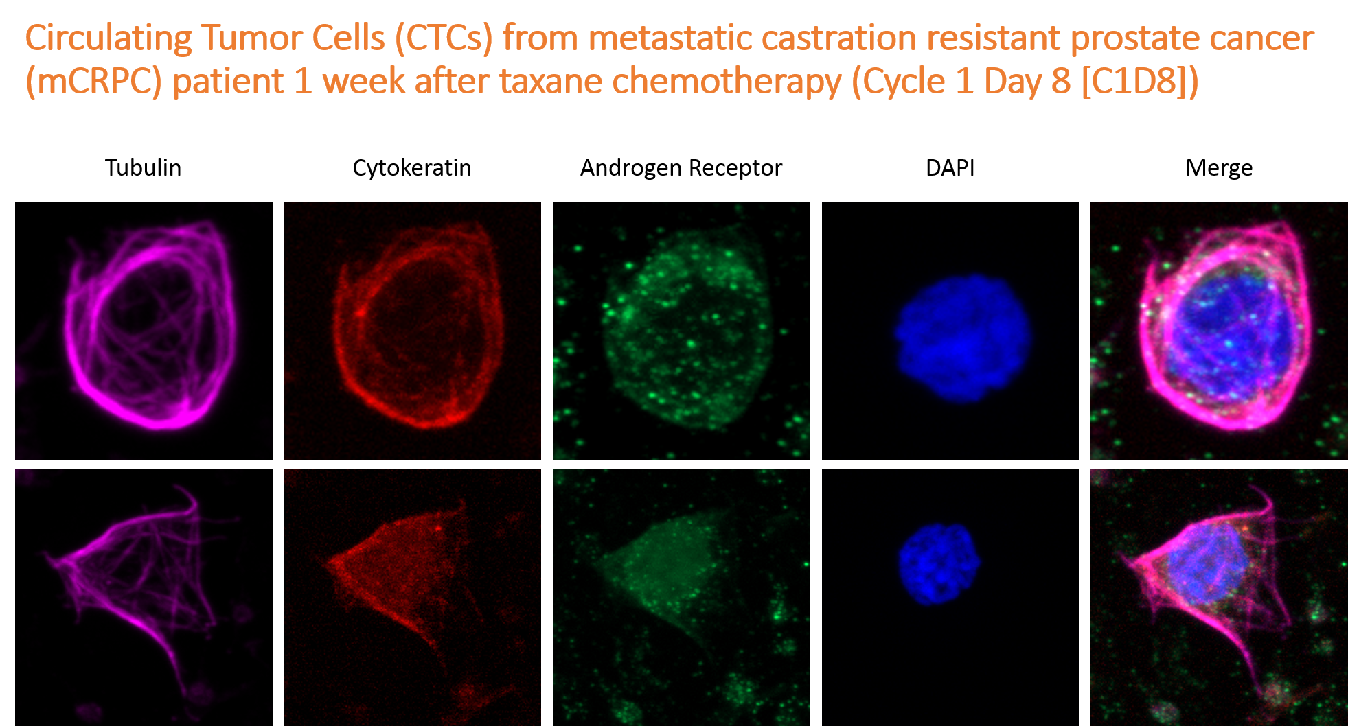
Circulating Tumor Cells (CTCs) post-treatment with taxane
Credit: David Worroll, Paraskevi Giannakakou (WCM)